See atlas 3.
Central laminated keratotic debris.
Keratin is a fibrous protein found in nails hair and the outer layer of skin.
Dna polymerase chain reaction pcr and gene sequencing studies performed on the tissue of the microscopic slide and paraffin block for the recently identified ts associated.
It is normally hard but can become soft under the nail in the presence of moisture.
Characteristics include generalized redness of the skin and severe.
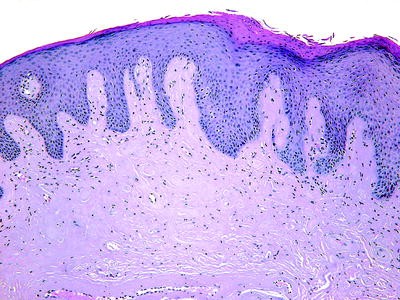
And a thinned variably ulcerated epidermis.
It usually occurs in the middle aged or elderly and is due to excessive exposure to the sun.
Electron microscopic studies of a follicular lesion showed extracellular viral particles suggestive of a polyomavirus within the central follicular keratotic debris.
On histologic examination the tumors are composed of an endophytic exophytic cup shaped squamous epidermal proliferation containing a crater like center filled with laminated keratotic material fig.
Right side contact us all pages.
A mixed inflammatory infiltrate.
Multiple keratoacanthomas may be seen in muir torre syndrome associated with sebaceous neoplasms and carcinomas of internal organs.
Hypertrophy of the cornea.
Nail disorders are often due to keratin debris spreading within the nail bed.
Subsequent re epithelization from the adjacent epidermis covers this entire process from the base.
Other links at bottom.
Epidermolytic hyperkeratosis a hereditary autosomal dominant form of ichthyosis present at birth.
Two pyogenic granulomas arising from the wall of an epidermoid cyst on the midback of a 62 year old white man are reported.
Keratosis ker ah to sis any horny growth such as a wart or callosity.
Left side donate all pages.
The dermal connective tissue inflammation and the keratotic debris degenerate to form the basophilic debris which corresponds to the keratotic plug.
A recent study described two siblings who developed kd at 2 and 4 years of age and were examined at 7 and 10 years of age respectively.
Actinic keratosis a sharply outlined wartlike or keratotic growth which may develop into a cutaneous horn and may become malignant.
This is exuded from the invagination seen in the fully evolved form of the lesion.
Called also senile or solar keratosis.
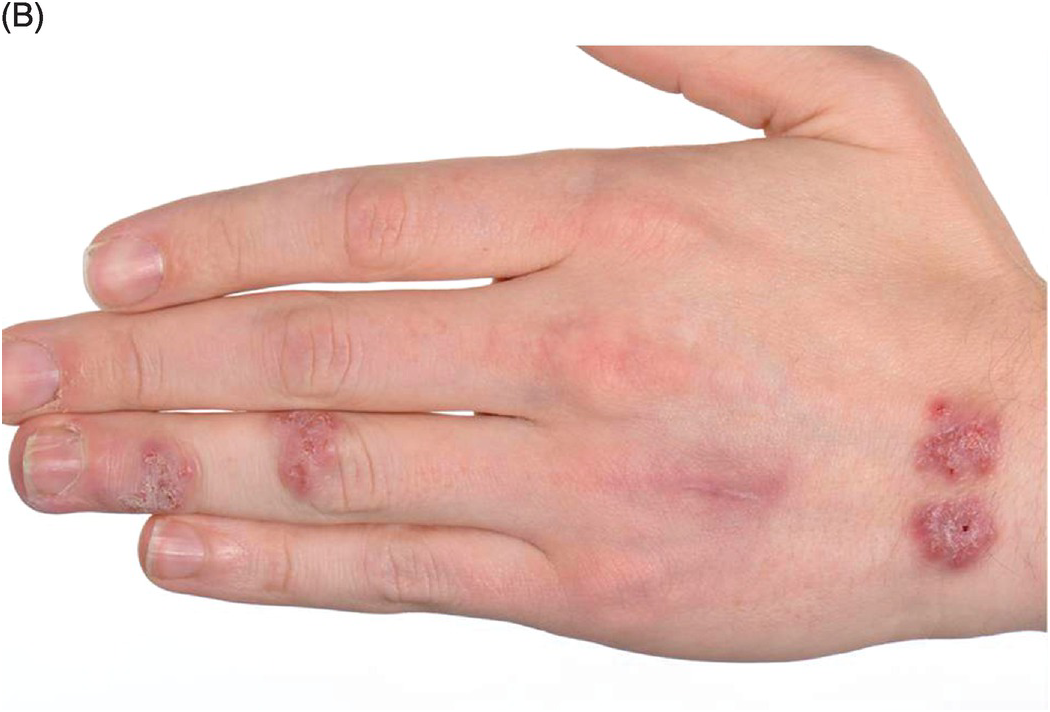
Presents with pruritic hyperkeratotic and ulcerated nodules and papules with a central keratotic plug mostly located on extensor surface of upper and lower limbs and on the trunk.
Keratotic debris ending with.
Hypertrophy of the horny layer of the skin or any disease characterized by it.
The lesions mainly occur on the scalp back and extensor surface of the legs and are asymmetrically distributed and hyperpigmented with central round or irregularly shaped keratotic plugs.